Among human organs the thymus stands out for giving up earliest. Its active tissue starts yielding to fat in childhood, and by the age of 50–60 only a small share of working epithelium is left. The process is called thymic involution, and it holds an unusual place in the biology of ageing: it is a rare case where the decline of an organ can be dated and measured. Below is what is known about it, how old that evidence is and what has been tried against it.
Involution starts earlier than people assume
For a long time it was pinned on sex hormones: puberty arrives, the thymus folds. A study by Steinmann and colleagues spoiled that logic. Morphometric analysis showed that the thymic epithelium shrinks steadily from early childhood and does not depend on puberty (Scand. J. Immunol., 1985). The organ begins folding long before a convenient hormonal explanation appears.
Palmer's review covers the quantitative side: the output of new T cells falls across the whole lifespan rather than collapsing at one moment (Frontiers in Immunology, 2013). The bone marrow changes in parallel, and together the two are described as ageing of the primary lymphoid organs (Seminars in Immunology, 2012). We covered the structure of the gland itself in a separate piece on the thymus gland.
What follows for immunity
The consequence is not that immunity becomes weak. It becomes different. Naive T cells able to answer an unfamiliar pathogen grow scarcer, and memory cells against things the body has already met take up the space. The repertoire narrows. Goronzy and Weyand examine the molecular mechanisms of that shift (Nature Reviews Immunology, 2019), Nikolich-Žugich describes the wider picture of immune ageing (Nature Immunology, 2017), and Thomas and colleagues tie thymic involution directly to immunosenescence and chronic inflammation (Immunity & Ageing, 2020).
Trying to regrow a thymus: the TRIIM study
The loudest modern attempt to intervene is the TRIIM study. Nine men aged 51–65 received growth hormone together with dehydroepiandrosterone and metformin for a year. The authors reported signs of thymic tissue recovery on MRI (magnetic resonance imaging) and a reduction in epigenetic age of roughly a year and a half across several DNA methylation clocks (Aging Cell, 2019).
Then comes the part popular retellings skip. There were nine participants. There was no control group at all. There was no blinding. Epigenetic age was not the primary endpoint, and the clock result came out of an additional analysis. The extended follow-up, TRIIM-X, recruited participants, but its results still have not appeared as a peer-reviewed paper. This is not a refuted study, it is an unreplicated one: an interesting signal nobody has independently checked.
The peptide line: what the Soviet school did
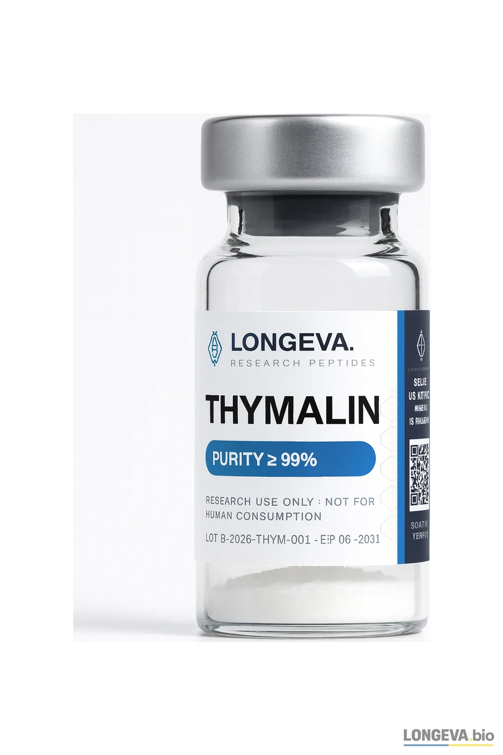
A separate body of thymus work grew up in the USSR. A polypeptide fraction known as thymalin was extracted from calf thymus and studied as an immunomodulator from the 1970s onward. A review by Morozov and Khavinson in an international journal describes the class of thymic peptides and the mechanistic thinking of the time (Int. J. Immunopharmacol., 1997), and a later review by Anisimov and Khavinson carries that logic into gerontology (Biogerontology, 2009).
This body of work deserves an honest reading. Most of the primary studies came out of a single scientific school, were published in Russian between the 1980s and the 2000s and are poorly accessible for independent checking. The clinical studies are mostly small and open-label, without placebo control. That does not make them wrong by default, but it does mean the level of evidence is not the level recommendations are built on. An independent 2024 review treats thymic peptides as a class and records the same heterogeneity (Int. J. Peptide Res. Ther., 2024). We examined the evidence for thymalin specifically in a separate piece on what the studies show.
It is worth noticing how differently the two stories are built. TRIIM is one small uncontrolled study waiting to be repeated. The peptide line is decades of publications that largely repeated themselves. Neither delivers what is usually wanted from them: a large independent trial with a control group.
What is linked to the thymus without any peptides
One finding deserves separate mention, because it is independent and comparatively easy to check. Duggal and colleagues compared amateur cyclists aged 55–79 with sedentary peers and found that several features of immunosenescence, including reduced thymic output of T cells, were far less pronounced in the physically active group (Aging Cell, 2018). This is an observational study, so it does not establish causation. On signal quality, though, it compares well against most of what gets sold as thymus restoration.
An honest bottom line
Thymic involution is a well documented fact with more than forty years of measurement behind it, and its link to immune ageing is described in current reviews. Human ability to reverse the process remains a hypothesis: there is one small uncontrolled study and a large body of old work from one school. If you see a text quoting a specific percentage of thymus restoration, there is almost certainly no study behind that number that would survive scrutiny.
Longeva supplies thymic peptides as reagents for research and educational use. They are not medicines, they are not intended to treat or prevent disease, and we make no claims of therapeutic effect.
Read also: Khavinson peptides (bioregulators): what they are and what research says.
See on Longeva
Research-use Thymalin with an open COA (certificate of analysis). Full overview in the wiki.